
显微镜核心设施
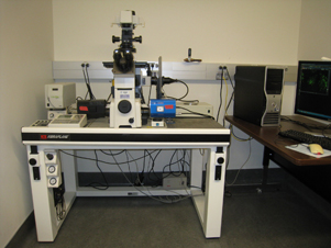
IMM显微镜核心在共聚焦显微镜,宽场荧光显微镜,Brightfield显微镜和图像分析方面提供了帮助。该设施配备了Leica TSC SP5直立的共聚焦显微镜,带有传统和共振扫描仪,尼康Eclipse TE2000E TE2000E倒置的宽视野显微镜,Zeiss Axio Scope Brightfield显微镜以及运行AMIRA软件的计算机工作站,以用于分析图像后数据分析成像数据的分析。。
The Microscopy Core will support the research needs of IMM research investigators by providing microscopy technical support, training and consultation.
Leica TCS SP5 Confocal Microscope

Leica TCS SP5直立共聚焦显微镜是带有串联扫描仪的点扫描共聚焦显微镜。谐振扫描仪可在512 x 512像素下以高达25 fps的帧速率实现高速成像,并且常规扫描仪允许在高达8192 x 8192像素(64兆像素)的框架尺寸下进行高分辨率成像。
关键功能包括:
- 激光照明线:氩458/476/488/514NM,DPSS 561NM,HENE 594NM和HENE 633NM。
- AOTF选择和激光线的衰减;能够进行感兴趣区域(ROI)扫描。
- Spectral detection in individually tunable wavelength bands.
- 三个用于荧光检测的光电倍增管(PMT),另一个用于发射光(明亮场,差分干扰对比度(DIC))。
- 软件染料分离功能。
- 针对共聚焦扫描优化的计划APO目标:10倍(NA 0.4)和20倍(Na 0.7)干燥,40倍(Na 1.25)和63倍(NA 1.4)油,以及63X(Na 1.2)水。63倍水物镜具有校正项圈。
关于共聚焦显微镜样品制备的注释:
- Our confocal microscope does not have a source to excite DAPI or Hoechst. Nucleic acid stains to be used with this microscope include but are not limited to: SYTOX Green, SYTOX Orange, CyTRAK Orange, Draq5, TO-PRO-3.
- 样品需要安装在适当的培养基中,并受到抗弹性试剂保护的荧光。
- Samples need to be covered with 0.17mm thick coverslips. The coverslip should be sealed securely to the slide/dish holding the sample.
- 为了确保您的样本将与共聚焦显微镜配合使用,潜在用户请在准备样本之前与服务中心经理讨论您的成像实验。
Nikon Eclipse TE2000E广场荧光显微镜
The Nikon Eclipse TE2000E inverted microscope provides for widefield fluorescence and transmitted light imaging. Fluorescence filter sets include DAPI, CFP, FITC, YFP, Texas Red, and Cy5.
关键功能包括:
- 高速先前的科学激发和排放过滤轮。
- 高灵敏度单色光学计量学级联512B EMCCD摄像头。
- 高分辨率单色光学量表冷却HQ2摄像头。
- 计划荧光4X(NA 0.13)目标。
- 计划荧光10倍(NA 0.3)和20倍(NA 0.45)长的工作距离目标,具有相位对比度,以通过塑料容器查看细胞。20倍物镜具有校正项圈。
- 计划APO 40X(NA 0.95)干燥和60倍(NA 1.4)的油目标,用于通过盖玻片查看样品。40X目标具有校正项圈和DIC棱镜。
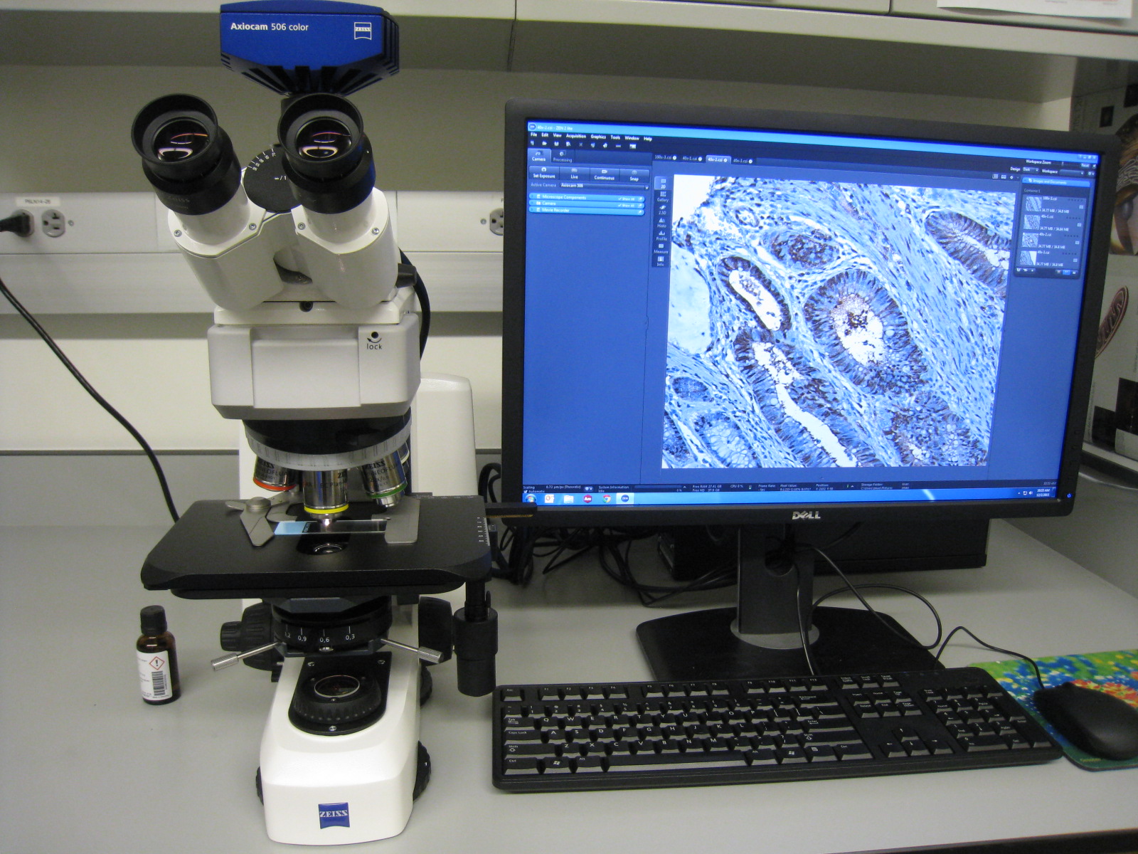
Zeiss Axio范围Brightfield显微镜
直立的Zeiss Axio范围为组织学和免疫组织化学样品的传输光亮场成像提供。
关键功能包括:
- AxioCam 506彩色相机。6百万像素颜色相机拍摄大型样品区域的图像。它提供的动态范围为1:2500,使最大的颜色差异和最小的细节在无损质量方面可见。Peltier冷却可确保低噪声和可重现的颜色图像质量。
- 可选的0.63倍适配器进一步允许相机拍摄超大样品区域的图像。
- EC Plan-Neofluar 5X(NA 0.16)目标。EC PLAN-neofluar 10X(NA 0.3)目标。EC Plan-Neofluar 20X(NA 0.5)目标。EC PLAN-NEEFLUAR 40X(NA 1.3)油目标。平面浓度100倍(NA 1.4)油目标。
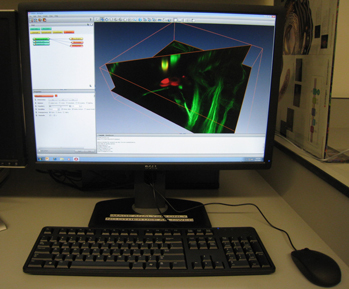
Image Analysis Workstation with Amira Software
一个专用的计算机工作站运行最新的version of Amira software and enables advanced visualization, analysis and presentation of imaging data. Users are welcome to bring images acquired from any microscope (in our facility or elsewhere). No previous experience with the software is required. Assistance is available upon request.
关键功能包括:
- 交互式3D导航
- 正交和倾斜切片
- 音量渲染
- 表面渲染
- 反卷积
- 分割
- Skeletonization and tracing of neural and vascular networks
- 图像注册
- 定量
- Co-localization analysis
- 多组分分析
- Animation tools and movie generation
Acknowledgements in publications demonstrate the value of our core and help us obtain financial and other support. So that we may continue to serve you in the best possible way, investigators who made use of our equipment and services should please acknowledge the IMM Microscopy Core in your publications and notify us of such acknowledgements.
Contacts:
Eva M. Zsigmond博士,导演
电话:713.500.2453 |电子邮件:Eva.M.Zsigmond@uth.tmc.edu
Zhengmei Mao博士,经理
Phone: 713.500.3389 | Email:zhengmei.mao@uth.tmc.edu
Uthealth
棕色基金会分子医学研究所
1825 Pressler Street SRB 616
Houston, Texas 77030